Cerebrospinal Fluid and Ventricular System Anatomy
## Cerebrospinal Fluid and Ventricular System Anatomy – Complete SEO-Friendly Guide
### SEO Title
**Cerebrospinal Fluid and Ventricular System Anatomy**
### Meta Description
Detailed anatomy of cerebrospinal fluid and the ventricular system covering formation, circulation, absorption, functions, ventricular components, and important clinical correlations.
### Keywords
cerebrospinal fluid anatomy, ventricular system brain, lateral ventricles anatomy, third ventricle anatomy, fourth ventricle anatomy, CSF circulation, choroid plexus, arachnoid villi, hydrocephalus anatomy
---
## 1. Cerebrospinal Fluid (CSF)
### Definition
Cerebrospinal fluid is a **clear, colorless fluid** that circulates within the **ventricular system of the brain and subarachnoid space** surrounding the brain and spinal cord, providing protection, nutrition, and waste removal.
### Normal Volume and Pressure
* Total volume (adult): **≈150 mL**
* Daily production: **≈500 mL**
* Normal opening pressure (lumbar puncture): **70–180 mm H₂O**
---
## 2. Formation of CSF
### Choroid Plexus
CSF is primarily produced by the **choroid plexus**, a vascular structure lined by **ependymal cells**.
**Locations of choroid plexus**
* Lateral ventricles (body and temporal horn)
* Third ventricle
* Fourth ventricle
**Mechanism**
* Active secretion via **Na⁺/K⁺ ATPase**
* Water follows osmotically
* Independent of intracranial pressure
---
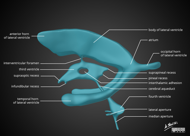
## 3. Ventricular System of the Brain
The ventricular system consists of **four interconnected cavities** lined by ependyma and filled with CSF.
---
### 3.1 Lateral Ventricles (First and Second Ventricles)
**Location**
* One in each cerebral hemisphere
**Parts**
1. **Anterior (frontal) horn**
* In frontal lobe
* Roof: Corpus callosum
* Floor: Head of caudate nucleus
2. **Body**
* Extends through parietal lobe
3. **Posterior (occipital) horn**
* In occipital lobe
4. **Inferior (temporal) horn**
* In temporal lobe
* Floor: Hippocampus
* Roof: Tail of caudate nucleus
**Communication**
* Each lateral ventricle communicates with the third ventricle via the **interventricular foramen (foramen of Monro)**
---
### 3.2 Third Ventricle
**Location**
* Midline cavity between the two thalami
**Boundaries**
* Lateral walls: Thalamus and hypothalamus
* Floor: Hypothalamus
* Roof: Tela choroidea
* Anterior wall: Lamina terminalis
* Posterior wall: Pineal region
**Connections**
* Receives CSF from lateral ventricles
* Drains into the fourth ventricle via the **cerebral aqueduct (aqueduct of Sylvius)**
---
### 3.3 Fourth Ventricle
**Location**
* Between pons and medulla anteriorly
* Cerebellum posteriorly
**Boundaries**
* Floor: Rhomboid fossa
* Roof: Superior and inferior medullary vela
**Openings**
* **One median aperture (foramen of Magendie)**
* **Two lateral apertures (foramina of Luschka)**
These openings allow CSF to enter the **subarachnoid space**.
---
## 4. Circulation of CSF
**Flow pathway**
1. Lateral ventricles
2. Foramen of Monro
3. Third ventricle
4. Cerebral aqueduct
5. Fourth ventricle
6. Foramen of Magendie and Luschka
7. Subarachnoid space
8. Arachnoid villi and granulations
9. Superior sagittal sinus
---
## 5. Absorption of CSF
### Arachnoid Villi and Granulations
* Protrusions of arachnoid mater into venous sinuses
* Act as **one-way valves**
* Absorption occurs when CSF pressure exceeds venous pressure
Minor absorption also occurs via:
* Spinal nerve sheaths
* Choroid plexus
---
## 6. Composition of CSF
* Clear and acellular
* Low protein
* Low potassium and calcium
* Higher chloride compared to plasma
* Glucose ≈ 60% of plasma glucose
---
## 7. Functions of CSF
* **Mechanical protection** (shock absorber)
* **Buoyancy** (reduces effective brain weight)
* **Nutrient delivery**
* **Removal of metabolic waste**
* **Maintenance of intracranial pressure**
---
## 8. Blood–CSF Barrier
Formed by:
* Tight junctions between **choroid plexus epithelial cells**
Functions:
* Regulates composition of CSF
* Protects CNS from toxins
---
## 9. Clinical Correlations
### Hydrocephalus
* Abnormal accumulation of CSF
**Types**
* **Non-communicating (obstructive):** Block within ventricular system (e.g., aqueductal stenosis)
* **Communicating:** Impaired absorption at arachnoid villi
* **Normal pressure hydrocephalus:** Triad of gait disturbance, dementia, urinary incontinence
---
### Raised Intracranial Pressure
* Headache
* Vomiting
* Papilledema
* Altered consciousness
---
### Lumbar Puncture
* Performed at **L3–L4 or L4–L5**
* Measures CSF pressure and composition
---
## 10. High-Yield Exam Points
* CSF production: **Choroid plexus**
* Narrowest part of ventricular system: **Cerebral aqueduct**
* Largest ventricles: **Lateral ventricles**
* Main absorption site: **Arachnoid granulations**
* CSF volume remains constant despite high daily production
---
If you want, I can also provide **MCQs (exam-oriented)**, **clinical case-based questions**, or **schema-style revision tables** for CSF and ventricles.